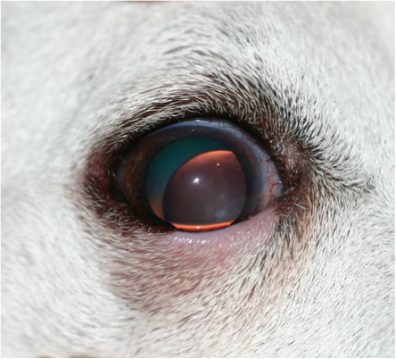
Cataracts

What is it?
Within the normal eye, the lens is used to help focus the incoming light onto the retina. It is similar to a camera lens. A cataract is an opacity or cloudiness of all or part of the lens. The lens is the fine focusing mechanism (approximately 25% of the refraction), the cornea is the course focusing mechanism (75% of the refraction).
Causes of Cataracts
The most common cause of cataracts in animals is an inherited defect in the lens. The breeding of related animals allows the abnormal genes to become concentrated and increases their chances for expression. Dogs, by far, are more greatly affected with genetic induced breed predisposed cataracts.
Other hereditary ocular diseases such as retinal degeneration (PRA), lens luxation, vitreous degeneration, and glaucoma are often associated with cataracts. In addition, cataracts can develop following injury, inflammation within the eye (iritis, uveitis), or from a systemic disease such as diabetes. In an older dog, the lens can develop into a cataract as a degenerative aging process (age-related cataracts).
It is normal for the lens to become slightly gray in color when dogs and cats become 8-10 years of age. This age change is associated with hardening of the lens known as nuclear sclerosis. The animal may have a difficult time focusing on close objects. This age related change begins in people at approximately 4-5 years of age. Hardening of the lens does not usually cause an appreciable loss of vision. Advanced hardening of the lens often progresses into senile cataract formation and decreased vision becomes apparent.
What are the possible complications of Cataracts?
Lens-Induced Uveitis (LIU)
During cataract formation, lens (cataract) proteins leak from the enclosed lens capsule and are recognized by the intraocular immune system as a foreign object. The identification of the leaking lens proteins stimulates an inflammatory reaction with the eye called lens induced uveitis (or cataract induced inflammation). Lens induced uveitis intensifies with chronicity, progression, and speed of onset of the cataract (i.e. hypermature and/or rapid onset cataracts typically have more severe lens induced uveitis). Uncontrolled lens induced uveitis can lead to secondary glaucoma, intraocular adhesions, corneal diseases, lens luxations, and retinal disease and detachments. Thus, it is very important to have this condition controlled prior to attempting surgery. Topical and/or oral anti-inflammatory medications will be administered for at least several days to a couple of weeks prior to cataract surgery. The more severe the lens induced uveitis, the more aggressive and longer the anti-inflammatory treatment. Patients who are not candidates for cataract extraction or who are not going to undergo cataract surgery should still be treated for this condition for as long as the cataracts remain.
Lens luxation and Subluxation
In this condition the lens becomes dislocated from its normal position. The ligaments (called zonules) which suspend the lens are ruptured and no longer hold the lens in its normal position. The displaced lens can be found in the front of the eye (anterior lens luxation), in the back of the eye (posterior lens luxation), or only partially displaced (lens subluxation). This condition can be primary (common in terriers and older cats) or can be secondary to other complicating ocular conditions such as glaucoma or uveitis. Primary luxated lens can induce secondary diseases including glaucoma, uveitis, and retinal or optic nerve diseases. Additionally, abnormal vitreous displacement into the pupil and front of the eye can be common in lens luxation / subluxation cases. These other ocular diseases would also concurrently need to be treated.
Secondary Glaucoma
Glaucoma occurs when the drainage angle becomes partially or completely obstructed and the aqueous being produced cannot exit properly causing pressure to build in the eye. If left untreated, this pressure can damage the retina and optic nerve leading to permanent blindness in as little as 24-48 hours. Glaucoma is characterized as either primary or secondary. Primary glaucoma is an inherited condition that occurs in many breeds including the Cocker Spaniel, Bassett Hound, Shar Pei, Chow Chow, Boston Terrier, Beagle and several other breeds. Primary glaucoma is often seen initially in one eye but eventually affects both eyes. Secondary glaucoma occurs as a result of another disease or trauma that causes the drainage angle to become blocked. Common causes of secondary glaucoma include trauma to the eye, inflammation, a luxated lens, intraocular hemorrhage and inflammation from cataracts.
Symptoms |
Common signs of ulcers can include:
-white opacity in the pupil of theye -decreasing vision -increased redness of the sclera (white part of the eye) |
Treatment |
The only effective treatment for cataracts is surgical removal. There have been many claims of nonsurgical cures by individuals since the beginning of time, but all medical treatments thus far have proven ineffective. Recently Ocu-Glo vision supplement (www.ocuglo.com) has shown promising research that it may slow the progression of cataracts, but will not reverse cataracts that are already present. Lasers have many uses in human and veterinary ophthalmology, however, lasers are not used to remove cataracts. Lasers are routinely used to treat certain complications that occur in humans after cataract surgery or other ophthalmic diseases such as glaucoma and retinal detachment. We use a laser for precataract surgery retinopexies.
Pre- surgical Health Protocol Cataract removal is a very intricate surgical procedure. A complete ophthalmic examination is necessary to determine if your pet is a candidate for cataract surgery. Your pet's general health is also very important. We require a preanesthetic blood work panel be performed within the 6 months prior to cataract surgery. A CBC and General Chemistry panel will make certain that the pet's internal organs (liver, kidneys, ect.) and blood counts are within normal limits. This preanesthetic blood work should be performed prior to the surgical date and can be completed by your regular veterinarian or at Veterinary Vision of Rochester. In the event of abnormalities, the doctor may recommend this be repeated the day of surgery. If the patient has other chronic or underlying health conditions, such as a heart murmur, liver disease or diabetes, additional testing may be recommended or required prior to anesthesia. Due to risk of post-operative infection, if your pet currently has dental disease, a dental cleaning will be required at least eight weeks prior to cataract surgery. Patients are not allowed to undergo bathing or grooming for 4 weeks after surgery, so it recommended that your pet be groomed a week or two prior to the scheduled surgery date. Pre-surgical Retinal Testing For cataract surgery to be successful, the entire eye, except for the cataractous lens, must be healthy. If the retina is not healthy, the eye can not receive light and send information to the brain (visual cortex) for interpretation. Often times, in our veterinary patients, the density of the cataract is too great and visualization and determination of retinal health is not possible. In these cases the electroretinogram (ERG) is an important pre-surgical test. It enables the doctor to test the viability of the retina. Cataract surgery will only be recommended if the ERG results are within acceptable limits. An ophthalmic ultrasound will also be recommended to evaluate the vitreous, lens and retinal for abnormalities that may predispose the patient a higher risk of post-operative complications such as retinal detachment. At our office both the ERG and ophthalmic ultrasound are mandatory tests before cataract surgery will be scheduled and must be performed within 4 weeks prior to surgery. Cataract Surgery The vast majority of patients will undergo a technique called phacoemulsification, a small incision, high frequency ultrasonic procedure. Intraocular Lens Implants (artificial lens) are placed in most patients. Typically, patients with an intraocular lens implant(s) have improved vision compared to patients without lens implants. Not every patient is a candidate for an intraocular lens implant, and animals without lens implants still have functional vision, but are far-sighted. Most patients show signs of vision immediately after surgery and this continues slowly over the next several weeks. Cataract surgery is typically an outpatient procedure. On the scheduled day of surgery, patients will be dropped off between 7 and 7:30 am. Cataract surgery typically lasts about 40-60 minutes per eye. Patients are then hospitalized for the day to monitor for post-operative complications. Barring any complications, patients are usually discharged between 3 and 5 pm. If complications are seen, the doctor may recommend your pet be hospitalized overnight for continued monitoring. Post Operative Period The post operative period is very important to a successful outcome. After surgery, there is an 8 week period of intensive home medical care. Patients are typically sent home on several topical medications 4-6 times daily, as well as oral antibiotics and anti-inflammatories. Due to the delicate sutures placed in the eye, an Elizabethan collar (cone) is required to be worn 24 hours a day for an average of 14 days following surgery. Self-trauma is the most common cause of incisional rupture, and will require additional surgery if allowed. Your pet may act strangely while adjusting to the collar as their peripheral vision is impaired by the collar. The will usually adjust in 24-48 hours. Patients are also kept on very restricted activity limits. Activities should exclude using a dog door, playing with toys, rough play and pulling on a leash. Some patients may require chemical sedation. Follow-up visits are very important in order to monitor the healing process . Most patients are examined at least 4 times during this 6-8 week period. If complications arise, additional recheck appointments may be necessary. Thereafter, your pet should have a 6-12 month follow-up ophthalmic exam. Potential complications of cataract development and/or surgery The vision, which is obstructed by the cataract, will be regained if the cataract can be removed without complications. If left untreated, the vision will most likely continue to digress until completely blind. Fortunately, cataract surgery is successful in most cases . Nevertheless, complications can occur in approximately 10% of cataract patients and a good result can not be guaranteed. Many eyes with cataracts have other abnormalities, which may not be evident at initial examination. Eyes with cataracts have a greater than normal risk for developing diseases including, but not limited to, retinal disease and detachment, glaucoma, corneal edema, intraocular hemorrhage, prolonged uveitis, intraocular adhesions, endophthalmitis and phthisis bulbi (shrinking of the eye). Cataracts will not return. Once a cataractous lens has been removed the cataracts will not regrow. After cataract surgery it is not uncommon to be able to see a small white membrane or film inside the pupil of the eye. This thin membrane is a portion of the lens capsule (like a sandwich baggie) that surrounded the cataract and serves as a barrier to maintain the vitreous in back of the eye. This is a very important function for the eye and it is not removed unless absolutely necessary. The white membrane is termed posterior capsular opacity (PCO) and in rare cases may extensively progress to limit vision. Diabetic patients and certain breeds may be at a slightly greater risk for of post-operative complications and may take longer to heal. Medical therapy and surgical procedures may be necessary to treat newly acquired disease as it occurs. At the time of you pet's initial ophthalmic examination, the doctor will advise you if your pet is a suitable candidate for cataract surgery and the potential complications. |
